Skulls Unlimited processes thousands of skulls yearly for hunters, professionals, and educators.
Learn more!

Our founder, Jay Villemarette, has been heard saying "Examining the similarities yet the differences between the species led me to become a lifelong collector." Now that your collection has expanded into multiple specimens of the same species, you can begin to piece together the life of the animal and how it may have differed from others in your library. Below we discuss three key areas to begin your comparative analysis and encourage you to dive deep into your own research for differences in your specimens.
3 Key Areas to Begin:
1. Pathology is a broad and generic term, but in this instance a companion item may help you distinguish abnormalities that lead to a diagnostic conclusion. Just like tissue shows bruises, bones show evidence of traumatic injuries that effected your specimen's life. In rare cases, you may find an instance of bone infections and metabolic bone disease.
2. Sexual dimorphism is the condition where the two sexes of the same species exhibit different characteristics beyond the differences in their sexual organs. In a large proportion of mammal species, males are larger than females. Differences may include size, weight, color, markings, and may also include behavioral and cognitive differences.
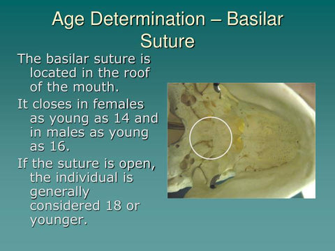
3. Age differences can also be determined in your comparative analysis. “Long in the tooth” is a common expression that stacks up! Fully erupted canine teeth and rear molars indicate an older specimen, while the presence of the basilar suture will tell you if you have an adolescent specimen. There are a variety of cranial sutures where their presence is typically found in young specimens, but the basilar sutures placement on the underside of the skull near the clivus. Well-defined ridges on or near connective tissue and tendons indicate full maturity, as well. Plenty of scholarly articles have been published on the subject of determining age from cranial sutures, if you want to take a deeper dive. See the images below for examples.

Source: Human Forensic Pathology Presentation Ch. 12
Warning: Images in this presentation are graphic.
0 of 3 items selected
Leave a comment